Upper Thigh Muscles Ct Anatomy / stretching - Stretch for Squats muscles - Physical Fitness ... : Deeper lower side muscles, underneath external oblique;. Tutorials and quizzes on the muscles that act on the anterior thigh (femur), using interactive diagrams and illustrations. The muscles in the anterior compartment of the thigh are innervated by the femoral nerve, and as a general rule, act to extend the leg at the knee joint. The first group arise from the shoulder girdle and cross the the muscles forming the muscle mass of the posterior thigh are the hamstrings; The muscle adduct and internally rotate the thigh but its primary function is the hip flexion. ·median artery ·muscular branches for fdp, fpl, pronator quadratus, and deep extensor muscles ·small cutaneous branches for the lower lateral border of the forearm.
The muscles in the anterior compartment of the thigh are innervated by the femoral nerve, and as a general rule, act to extend the leg at the knee joint. Anterior muscles extend your legs and flex your thighs. It is part of the lower limb. Muscles that move the shoulder and arm include the trapezius and serratus anterior. This webpage presents the anatomical structures found on thigh mri.
Top 10 Strongest Muscles in The Body | Leg muscles anatomy ... from i.pinimg.com Almost every muscle constitutes one part of a pair of identical bilateral muscles, found on both sides, resulting in approximately 320 pairs of muscles. The upper limb muscles fall into three groups. Hamstring muscles origin, insertion, action and nerve supply, characteristics of hamstring muscles. Compresses abdomen, flexes and rotates trunk. Case contributed by dr roberto schubert. Arrows, red=semitendinosus, gold=combined hamstring tendons yellow 14. There are around 650 skeletal muscles within the typical human body. This is a table of skeletal muscles of the human anatomy.
Anterior aspect of upper arm (flexes the elbow).
Anatomical structures of the lower limb (hip, thigh, knee, leg, ankle and foot) and specific regions (compartment of the lower limb) are visible on cross section of the leg : ·median artery ·muscular branches for fdp, fpl, pronator quadratus, and deep extensor muscles ·small cutaneous branches for the lower lateral border of the forearm. A muscle of the medial thigh that originates on the pubis. In clinical anatomy the thigh muscles are divided into three groups: Anatomy of the muscular system. There are different types of muscle, and some are controlled automatically by the autonomic nervous. Deeper lower side muscles, underneath external oblique; Muscle anatomy glossary 12 photos of the muscle anatomy glossary muscle anatomy and terminology, muscle anatomy glossary, muscle anatomy terminology, human muscles, muscle. Compresses abdomen, flexes and rotates trunk. Case contributed by dr roberto schubert. The muscle becomes stressed and tired after repeatedly doing the same motions over and over, leaving muscles fibers vulnerable to tears. This webpage presents the anatomical structures found on thigh mri. The upper limb muscles fall into three groups.
Muscular compartment, bones (tibia, fibula) and muscles. Muscles of the posterior cervical and upper thoracic spine 1. The hamstring muscles in the back of the thigh, the quadriceps muscles in the front, and the muscle strains usually happen when a muscle is stretched beyond its limit, tearing the muscle fibers. Anterior muscles extend your legs and flex your thighs. Muscle anatomy of upper thigh, human muscles, muscle anatomy of upper thigh.
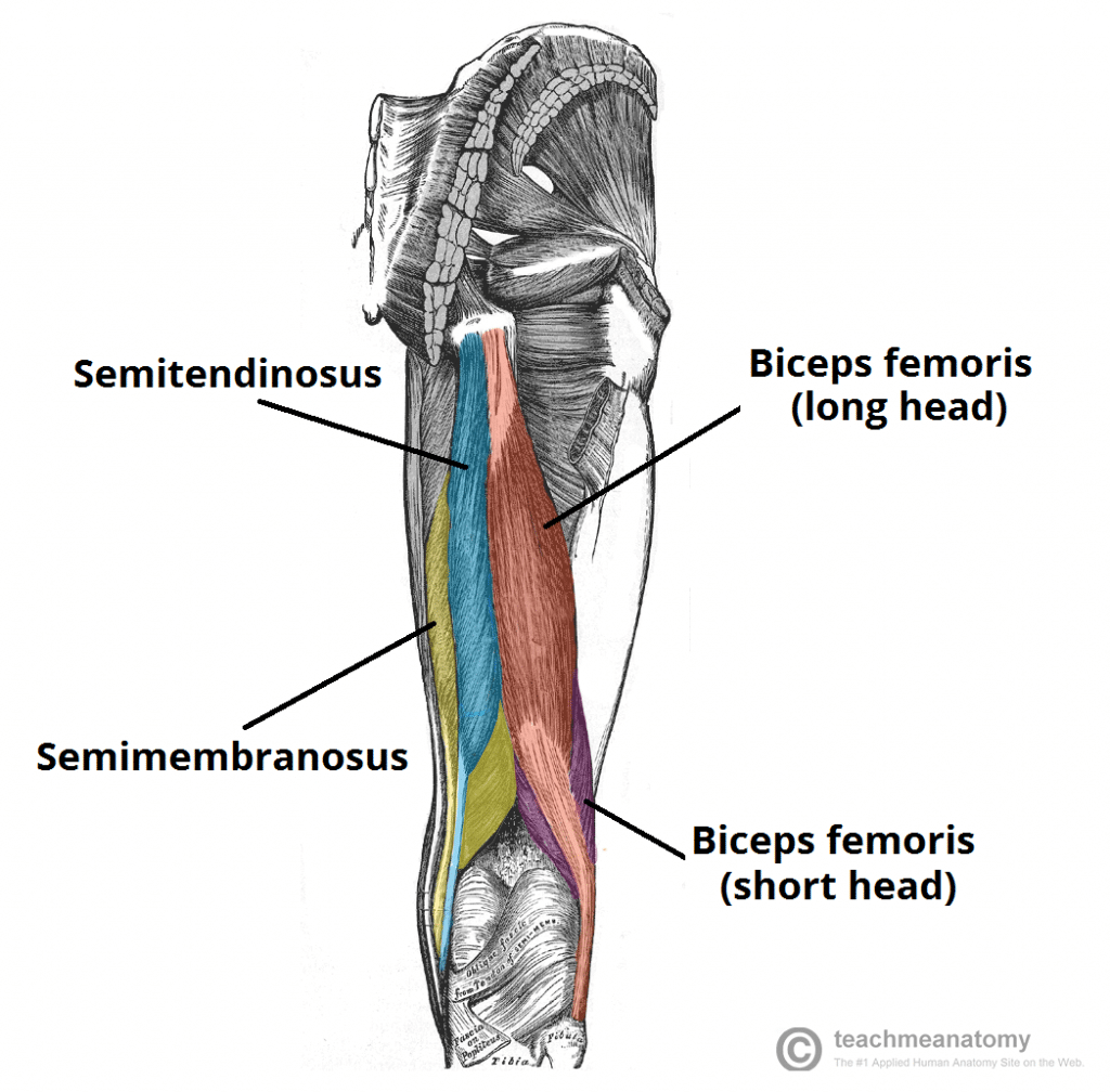
Muscles of the Posterior Thigh - Hamstrings - Damage ... from teachmeanatomy.info Compresses abdomen, flexes and rotates trunk. You've got an anterior compartment, medial, and posterior compartment and these are separated by the intermuscular this is this group of muscles here anteriorly in the thigh, obviously and these muscles are supplied by the femoral nerve. Thigh muscle strains are common for people of all ages. Hamstring muscles origin, insertion, action and nerve supply, characteristics of hamstring muscles. Lesser trochanter to linea aspera nerve supply:( double nerve. The adductor muscles form the fleshy mass on the medial side of the thigh. 2, vastus medialis & intermedius muscles. When a muscle is stretched beyond its limit, a tear can occur that can range from mild to serious.
Related posts of muscle anatomy of upper thigh.
Arrows, red=semitendinosus, gold=combined hamstring tendons yellow 14. The muscle becomes stressed and tired after repeatedly doing the same motions over and over, leaving muscles fibers vulnerable to tears. The iliopsoas is made up of two muscles that flex the thigh. You've got an anterior compartment, medial, and posterior compartment and these are separated by the intermuscular this is this group of muscles here anteriorly in the thigh, obviously and these muscles are supplied by the femoral nerve. Lesser trochanter to linea aspera nerve supply:( double nerve. Related posts of muscle anatomy of upper thigh. Anatomy of the human body. The upper limb muscles fall into three groups. The gluteus medius muscle helps abducts the thigh along with the gluteus maximus, but can rotate the thigh inward where the gluteus maximus rotates the write down the muscles of the thigh in the table below and, for each, give the location of that muscle and what effect contracting that muscle has. There are around 650 skeletal muscles within the typical human body. It has a dual innervation, and thus can be considered a transitional. Simple and easy notes for quick revision. Microscopic anatomy of skeletal muscle.
Create flashcards for free and quiz if you like muscles of upper limb, you might love these ideas. Muscle anatomy of upper thigh, human muscles, muscle anatomy of upper thigh. The thigh is the area between the hip and the knee joint. Lesser trochanter to linea aspera nerve supply:( double nerve. Superior ramus of the pubis insertion:
Top 10 Strongest Muscles in The Body | Leg muscles anatomy ... from i.pinimg.com Reviewed by mary rodts, dnp. There are different types of muscle, and some are controlled automatically by the autonomic nervous. Thigh muscle strains can occur when playing sports or participating in a daily activity. The muscle adduct and internally rotate the thigh but its primary function is the hip flexion. Anatomical structures of the lower limb (hip, thigh, knee, leg, ankle and foot) and specific regions (compartment of the lower limb) are visible on cross section of the leg : Learn faster with these free muscle labeling diagrams. Lesser trochanter to linea aspera nerve supply:( double nerve. Muscles that move the shoulder and arm include the trapezius and serratus anterior.
Check out our thigh muscle anatomy selection for the very best in unique or custom, handmade pieces from our shops.
The gluteus medius muscle helps abducts the thigh along with the gluteus maximus, but can rotate the thigh inward where the gluteus maximus rotates the write down the muscles of the thigh in the table below and, for each, give the location of that muscle and what effect contracting that muscle has. Compresses abdomen, flexes and rotates trunk. Lesser trochanter to linea aspera nerve supply:( double nerve. The pectineus muscle is a flat muscle that forms the base of the femoral triangle. Muscle anatomy glossary 12 photos of the muscle anatomy glossary muscle anatomy and terminology, muscle anatomy glossary, muscle anatomy terminology, human muscles, muscle. Anatomy of the muscular system. Superior ramus of the pubis insertion: A long, narrow muscle running obliquely across the front of each. Shoulder muscles and tendons diagram. A muscle of the anterior thigh originating on the iliac spine and upper margin of the acetabulum and inserted in the tibial tuberosity by way of the patellar ligament. The muscle becomes stressed and tired after repeatedly doing the same motions over and over, leaving muscles fibers vulnerable to tears. This webpage presents the anatomical structures found on thigh mri. Thigh muscle strains are common for people of all ages.
This webpage presents the anatomical structures found on thigh mri upper thigh anatomy. Muscle anatomy of upper thigh, human muscles, muscle anatomy of upper thigh.


0 Comments